The projects undertaken in CEBC cover a wide range in the field of Biomedical Engineering, involving both engineering and clinical students:
| Project Type | IIB project (2020 - 2021 academic year) |
| Student | Amy Edwards (Engineering Department) |
| Supervisor |
Prof Michael Sutcliffe (Engineering Department) |
| Collaborators |
Prof Paul White (Addenbrooke's Hospital), Stephen Large (Royal Papworth) |
Introduction
Heart transplantation is an excellent treatment for carefully selected patients with advanced heart failure. The deficit between patients in need of cardiac transplantation and the number of donor hearts is a serious problem, with almost 20% of listed patients dying whilst waiting for a heart transplant [1]. Donations after circulatory death (DCD), have the potential to significantly increase the number of overall donor hearts. However, because heart function cannot easily be assessed after circulatory death, DCD hearts carry extra complexity. The overall aim of this project is to develop a procedure to assess the functional status of the heart, and this MEng project reports on the first steps in this process.
Methodology
Figure 1 provides an overview of the approach taken. A balloon model is used to replicate most of the key factors present in the heart, including developing a pressure-volume (P-V) loop and wall motion. Missing is the active muscle function of the heart. A silicone mould phantom is used to enhance the compliance match with a heart, also providing options for systematic investigation of geometric and materials factors.
The experimental measurement set-up is illustrated in Figure 2. At the heart of the measurement system is an ADXL313 accelerator attached to the balloon wall. An experimental pumping rig has been developed to carry out inflation tests on the balloon and silicone models. The aim of the inflation test is to change the state of the deformations present in the balloon wall and to determine if these deformations can be characterised through accelerometery. This rig was used to measure the pressure and volume of the model to enable the effectiveness of the model as an analogue of the heart to be assessed. The displacements of the balloon walls during inflation and deflation are tracked using the chosen accelerometer. An image processing algorithm was used to validate the displacement measurements obtained from the accelerometer.
Results
Through the comparison of pressure volume data from the balloon model to that of a real heart, it is concluded that the balloon and silicone models can be used as an engineering analogue of the heart. Accelerometery is successful in characterising the displacements of the balloon wall which range from 5-15 mm with an average error of 33%. In a range of 0-50 mm the average error was 14% highlighting that the error increases as the scale of the displacements decrease.
Further work is needed to apply the system to silicone phantoms and a porcine heart, optimising the approach and assessing its efficacy, before making recommendations for use in humans.
Acknowledgements
Thanks to Nan Li, Alex Casabuena and Luis Andrade Acosta for their help with obtaining equipment and carrying out experiments.
References
- S. Messer, J. Lannon, E. Wong, C. Hopkinson, S. Fielding, R. Axell, A. Ali, S. Tsui, S. Large, The potential of transplanting hearts from donation after circulatory determined death (dcd) donors within the United Kingdom, The Journal of Heart and Lung Transplantation, 34(4), S275, 2015
* For more details please contact Prof Michael Sutcliffe
| Project Type | IIB project (2020 - 2021 academic year) |
| Student | Rosie Earl (Engineering Department) |
| Supervisor |
Prof Michael Sutcliffe (Engineering Department) |
| Collaborator |
Stanisław Tomaszczyk (Engineering Department), Dr Joel Melton (Addenbrooke's Hospital) |
Introduction
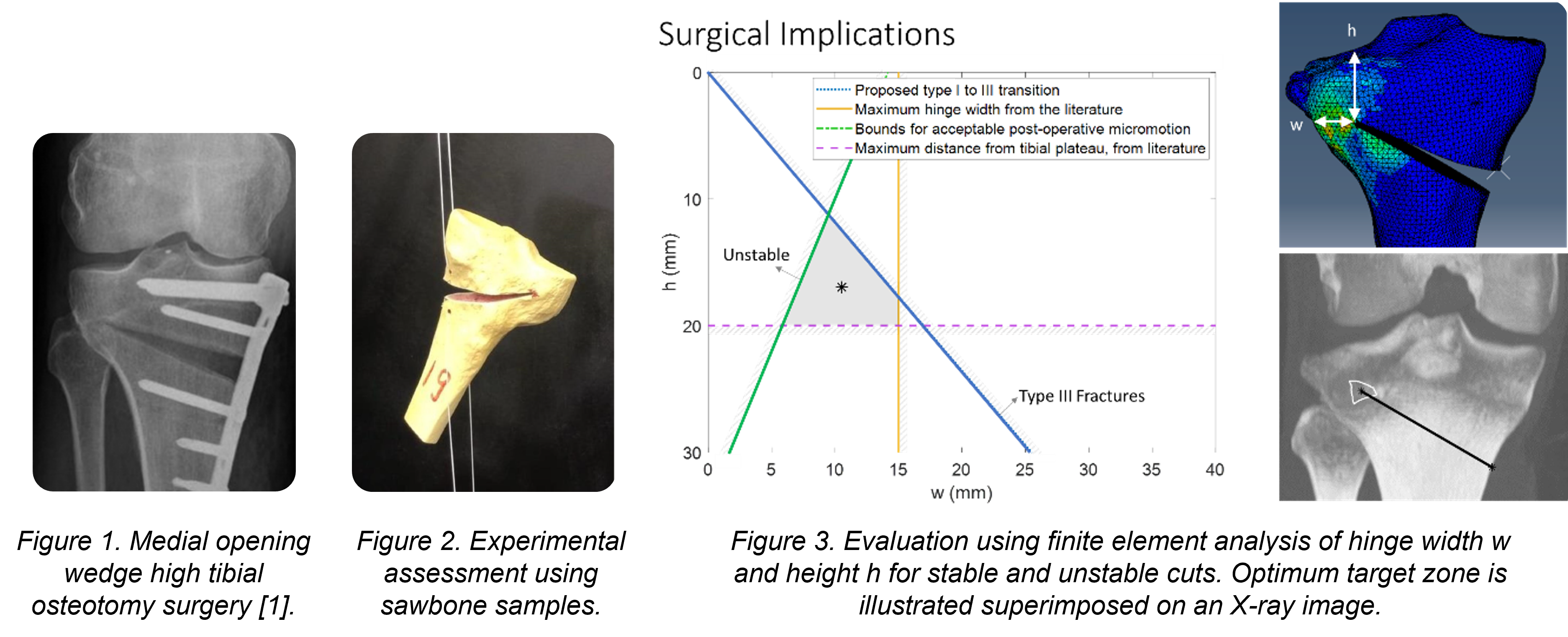
Medial opening wedge high tibial osteotomy (MOWHTO) surgery corrects angular misalignment around the knee with the aim of improving osteoarthritis. A cut is made part-way through the tibia bone and is opened on the medial side to form a wedge. This alters the leg alignment, correcting the location of the weight-bearing axis within the knee joint. The osteotomy is finally fixed in place with a plate and screws to allow for bone regrowth within the wedge. A radiograph of a fixed osteotomy is shown in Figure 1. The surgery leaves a ‘hinge’ of remaining bone on the lateral side, which is crucial for post-operative stability. However, a possible complication is fracture in this region, either during the wedge opening process or due to gait loading post-operatively. The aim of this study is to find the optimum cut geometry which minimises the chance and severity of lateral hinge fracture in MOWHTO surgery.
Methodology
An experimental study using artificial sawbone and perspex plate samples explored failure with different cut lengths and apex positions. Figure 2 illustrates the set-up for the sawbone sample, with wedge opening simulated within an Instron machine by applying loads using cord. The failure type and failure load was recorded, following the failure type terminology introduced by Nakamura et al [2].
A numerical study used a finite element (FE) model, as illustrated in Fig. 3, to assess the effect of hinge position on the stresses and failure mode both intra- and post-operatively.
Results
Both sets of experiments showed a clear decrease in the wedge opening angle at failure and increase in the failure load as the width of the lateral hinge got larger. Additionally, it was found that type I fractures are recorded for narrower hinges while type III failure was seen for wider hinges.
The results from the intra-operative simulations indicated that the stresses become larger as the hinge width increases, and the type of fracture changed from type I to the more severe type III. Post-operative stability of the osteotomy was quantified using the relative movement between the surfaces of the wedge. The FE results for intra- operative and post-operative loading conditions were combined with guidance from the literature to propose a region within the bone to place the osteotomy cut apex, as illustrated in Fig. 3.
Conclusions
Finite element modelling, supported by experiment observations, provides guidelines for the location of the cut apex for MOWHTO based on intra- and post-operative loading.
Acknowledgements
Thanks to Graham Treece for help with the imaging.
References
- P. Niemeyer et al., “Two-Year Results of Open-Wedge High Tibial Osteotomy With Fixation by Medial Plate Fixator for Medial Compartment Arthritis With Varus Malalignment of the Knee,” Arthroscopy - Journal of Arthroscopic and Related Surgery, vol. 24, no. 7, pp. 796–804, Jul. 2008, doi: 10.1016/j.arthro.2008.02.016.
- R. Nakamura et al., “Appropriate hinge position for prevention of unstable lateral hinge fracture in open wedge high tibial osteotomy,” The Bone and Joint Journal, vol. 99, no. 10, pp. 1313–1331, 2017, doi: 10.1302/0301-620X.99B10.
* For more details please contact Prof Michael Sutcliffe
| Project Type | IIB project (2020 - 2021 academic year) |
| Student | Filsan Hassan (Engineering Department) |
| Supervisor |
Dr Nan Li (Engineering Department) |
| Collaborator |
Dr Stephen Smith (Addenbrooke's Hospital) |
Introduction
Digital dermoscopes are imaging devices used by clinicians to inspect skin close-up. As well as being expensive tools, their efficacy is usually reliant on the expertise of the user to make a correct diagnosis by eye. Efforts have been made in the dermoscopy market to embed machine learning and optical imaging techniques to aid in the detection and interpretation of skin lesions. This project aims to implement the optical imaging technique of polarimetry which, although heavily researched, is not currently implemented in dermoscopes on the market. It is possible to infer significant information on the morphology of cells, e.g., cell size, orientation, and tissue margins which are important in distinguishing between malignant and healthy tissue, from polarimetric parameters.
Methodology
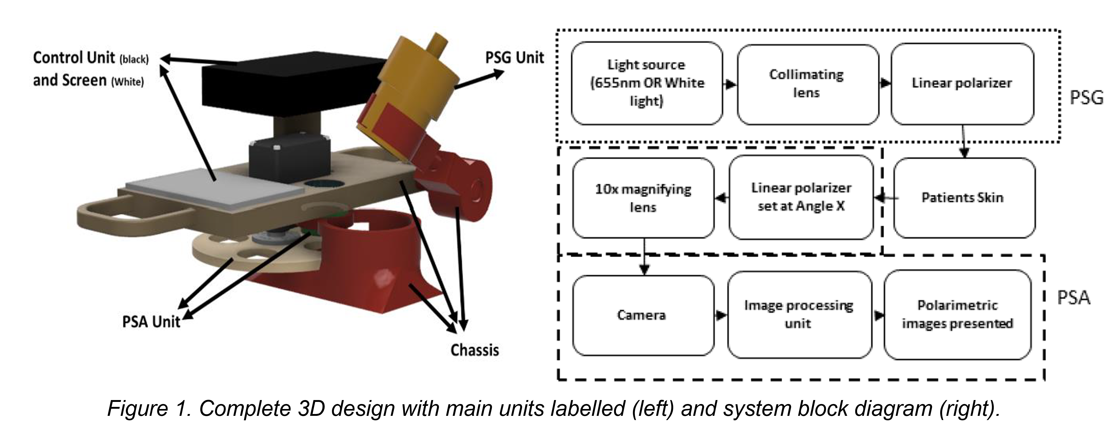
Affordable dermoscopic imaging will be offered by building the device from a Raspberry Pi and camera. Similarly, affordable polarimetric imaging will be provided by applying the method of division of time polarimetry (DoTP) and creating the necessary units from 3D printed parts and other low-cost materials (Figure 1). Furthermore, imaging with non-polarised white light should be embedded to take standard dermoscopic images and the additional imaging states of linearly polarised red (655nm) and linearly polarised white light imaging will be supported to measure important polarimetric parameters. These include cross-polarisation intensity and Stokes vector parameters which are specific to the evaluation of relevant surface and subsurface features. These measurements are translated to the user through appropriate colourmap images of the original lesions.
Image pre-processing, segmentation and registration algorithms based on computer vision techniques will be applied to the raw images to increase the accuracy of the classification parameters. The image pre-processing and segmentation algorithms enable lesion detection via Canny Edge detection and Otsu's thresholding, whilst the performance of two registration methods (Speeded Up Robust Feature (SURF) detection and Phase Correlation) will be applied and compared. Moreover, the lesion detection algorithm will also be used to define relevant geometric parameters of the lesion. The dataset of lesions imaged to assess these algorithms consist of 10 secondary malignant moles and three primary benign lesions.
Results
Overall, the dermoscopic images captured by the device have met several key standards set by the international skin imaging collaboration (ISIC) resulting in a suitable proposal for a low-cost remote dermascope once white light imaging is fully supported. Likewise, the three imaging states used to produce these images were calibrated to produce images of sufficient (ISIC) resolution. Imaging using red light, however, was found to obscure features from red-pigmented lesions whilst polarised light was found to obscure or accentuate some surface features. Therefore, it is recommended to only used white light images to make direct visual assessments of the lesions.
Furthermore, this change in structural features apparent across images at different states resulted in additional image registration constraints. The method of phase correlation was found to only be robust against changes in the polarisation state of white light whilst the ability to form correspondence from SURF was significantly hindered for polarised white and red-light images, especially for symmetrical lesions. A method of combatting this was derived by combining the lesion detection algorithm, which could identify boundaries for all imaging states, with the SURF feature detection algorithm. This produced enough correspondences to define the transformation and correct for misregistration in this dataset. The overall performance of this method and other registration methods applied to polarised images remain undefined due to the difficulty found in measuring similarity between dissimilar images.
The polarimetric parameters defined after registration produced the expected morphological relationships for benign lesions. The stokes vector images produced showed more susceptibility to misregistration than cross polarisation results. Furthermore, the geometrical parameters determined for malignant and benign lesions showed appropriate differences and the accuracy of these parameters was improved when Otsu's thresholding was applied.
Conclusions
The algorithms developed have been found to produce the expected results for images of healthy lesions. Furthermore, it has been found that these low-cost materials can adequately produce dermoscopic images and it is recommended that only the non-polarised white light images be used to make unaided visual assessment. The polarimetric imaging units developed function well separately suggesting that the addition of polarimetric imaging in dermoscopes is both plausible and valuable. Future work should focus on miniaturising and improving the integration of the units developed. Likewise, the algorithms have not been applied to lesions affected by melanoma therefore future work should complete clinical testing to deduce the validity of these algorithms and hence develop these further if required.
* For more details please contact Dr Nan Li
| Project Type | IIB project (2020 - 2021 academic year) |
| Student | Savannah Dixon (Engineering Department) |
| Supervisor |
Dr Nan Li (Engineering Department) |
| Collaborator |
Abigail Bush, Alice Tait (Addenbrooke's Hospital) |
Introduction
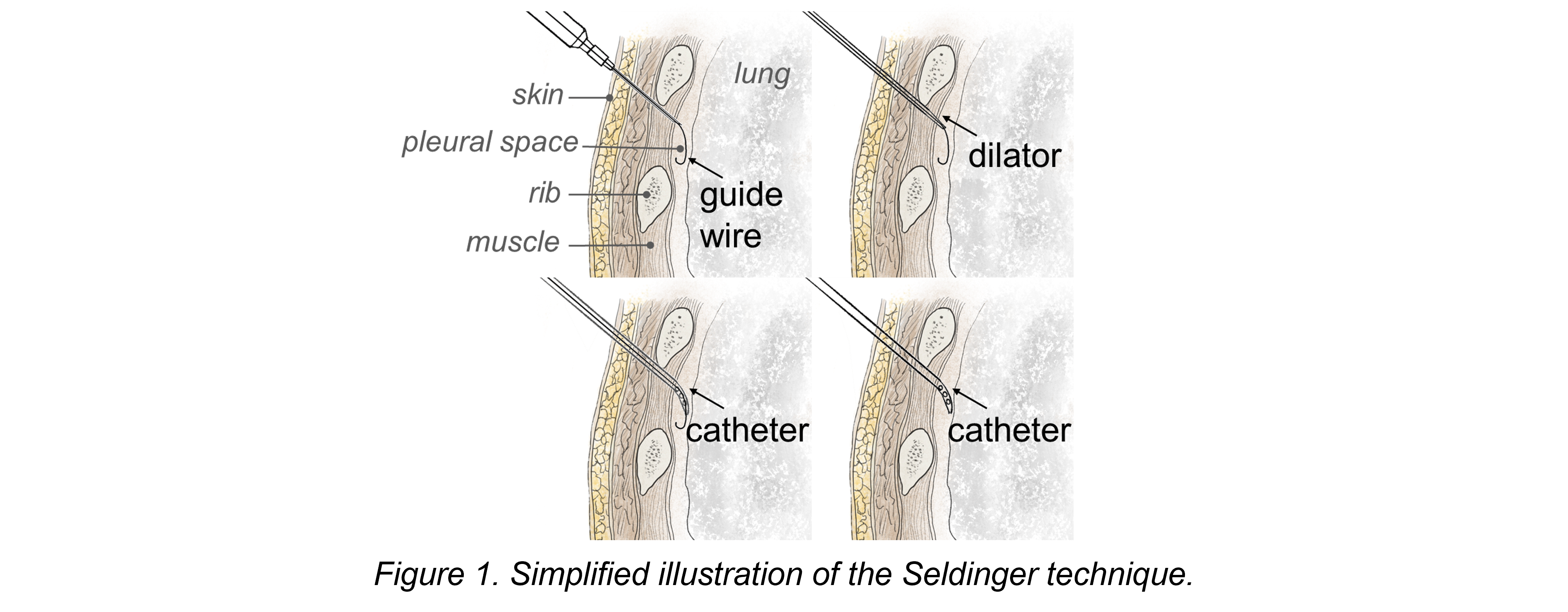
Pneumothorax is a condition where air builds up in the pleural cavity, the space between the lung and the chest wall. This disrupts the pressure that keeps the lungs inflated. The incidence of pneumothorax is higher in neonates than in any other age bracket [1]. The Seldinger technique (Figure 1) is the suggested procedure to treat a pneumothorax in a non-emergency situation by NHS [2]. Control over the insertion procedure requires extremely high dexterity, especially in neonates. The current technique has little to no feedback to communicate where the device is positioned in the chest wall. This can result in life-threatening complications, primarily over-introduction of the device into the chest cavity causing organ perforation. Often this mistake is not immediately detectable in the patient’s condition [3] and further complications can occur such as catheter blocking or kinking. This project aims to explore alternative, cost effective methods for chest drain insertion in neonates that provides sufficient control and feedback to eliminate the risk of organ perforation.
Methods
This project adopts both experimental and observational studies [4] for the device design and scrutinised clinical assessment. The methodology has focused on the application of a systems based, user focused design methodology as described in the Healthcare Design Toolkit [5]. The success of the alternative device designs and methods is assessed through the clinical experience of control and feedback in a simulated setting. In close collaboration with clinical staff, a series of ‘works like’ prototypes were developed iteratively for the final trial.
Results and Discussion
The prototypes were evaluated in a simulation with multiple staff members led by Dr Rajiv Chaudhary. This provided deep insight into how the development of novel technology could support the goal of improving clinical practice. The simulation results indicated that a number of the mechanisms developed and trialled have the potential for delivering a step change in improving the safety of this procedure for neonates.
Conclusions
A number of mechanisms for chest drain insertion are promising in terms of both impacts on safety and feasibility. Steps for further work will be outlined.
Acknowledgements
Dr Rajiv Chaudhary, Cambridge University Hospitals NHS Foundation Trust
References
- H. Dahmarde et al., "Accuracy of ultrasound in diagnosis of pneumothorax: a comparison between neonates and adults—a systematic review and meta-analysis." Canadian respiratory journal 2019 (2019).
- R. Chaudhary et al., Clinical guideline: Insertion of intrapleural chest drain 2021 (in progress).
- RC. Reed et al., "Complications of percutaneous thoracostomy in neonates and infants." Journal of Perinatology 36.4 (2016): 296-299.
- A. Taktak et al., eds. Clinical engineering: a handbook for clinical and biomedical engineers. Academic Press, 2019.
- PJ. Clarkson et al., "Engineering better care: a systems approach to health and care design and continuous improvement." (2017): 1-92.
* For more details please contact Dr Nan Li
|
Project Type |
IIB project (2020 - 2021 academic year) |
|
Student |
Amir Fariq bin Anuar (Engineering Department) |
|
Supervisor |
Dr Nan Li (Engineering Department) |
Introduction
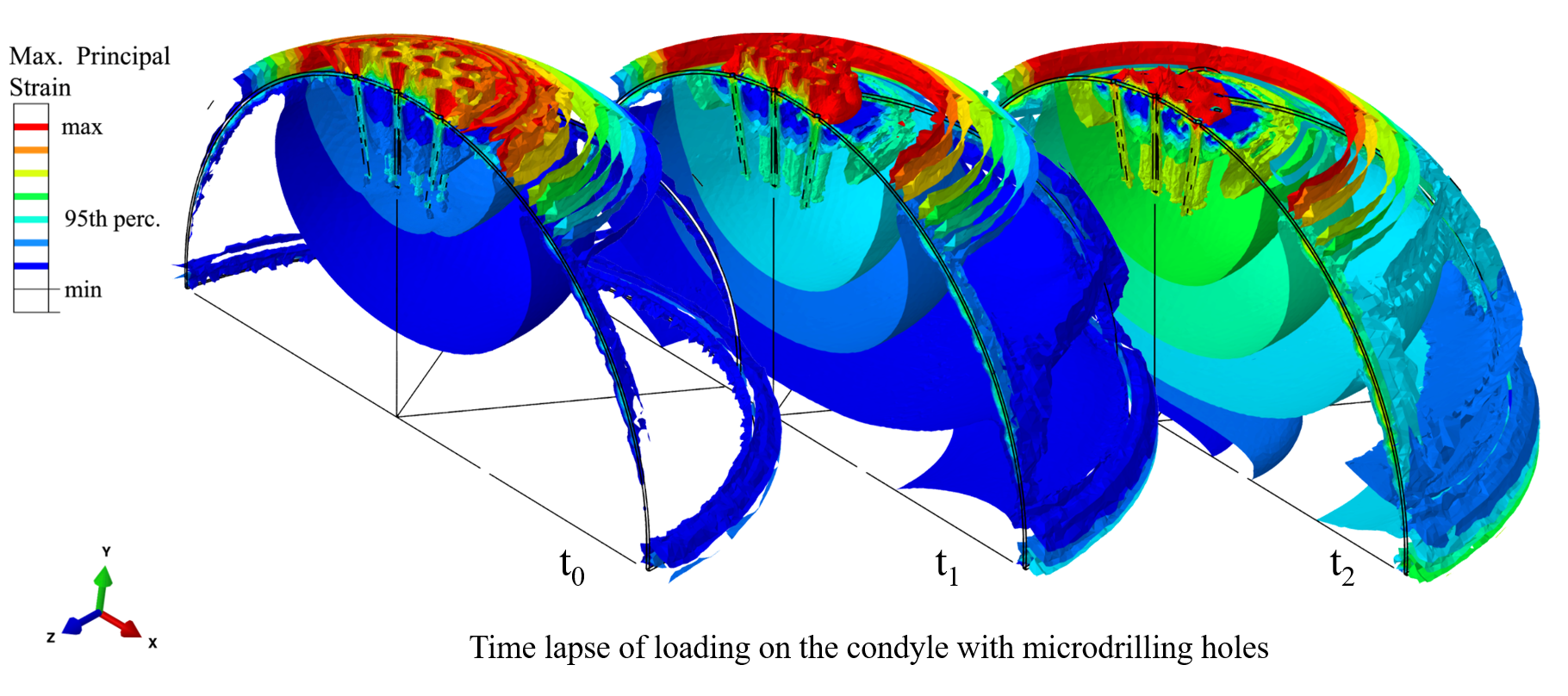
Microdrilling is a marrow stimulation technique that aims to treat osteoarthritis and other cartilage defects by drilling holes in the bone to induce bleeding and healing [1]. Microdrilling is a relatively simple cartilage repair technique with technical ease, does not require advanced tools or biological material, and represents a crucial early-intervention technique that has the potential to prevent total joint arthroplasty. However, the effects of various parameters of microdrilling are less understood, and the application of microdrilling is not standardised.
Literature review was done to investigate the current state of understanding of microdrilling and find typical values of drilling parameters and their effects. It was found that arrangement of holes [2], more specifically, hole coverage area [3], has not been thoroughly investigated, understood nor standardised.
Methodology
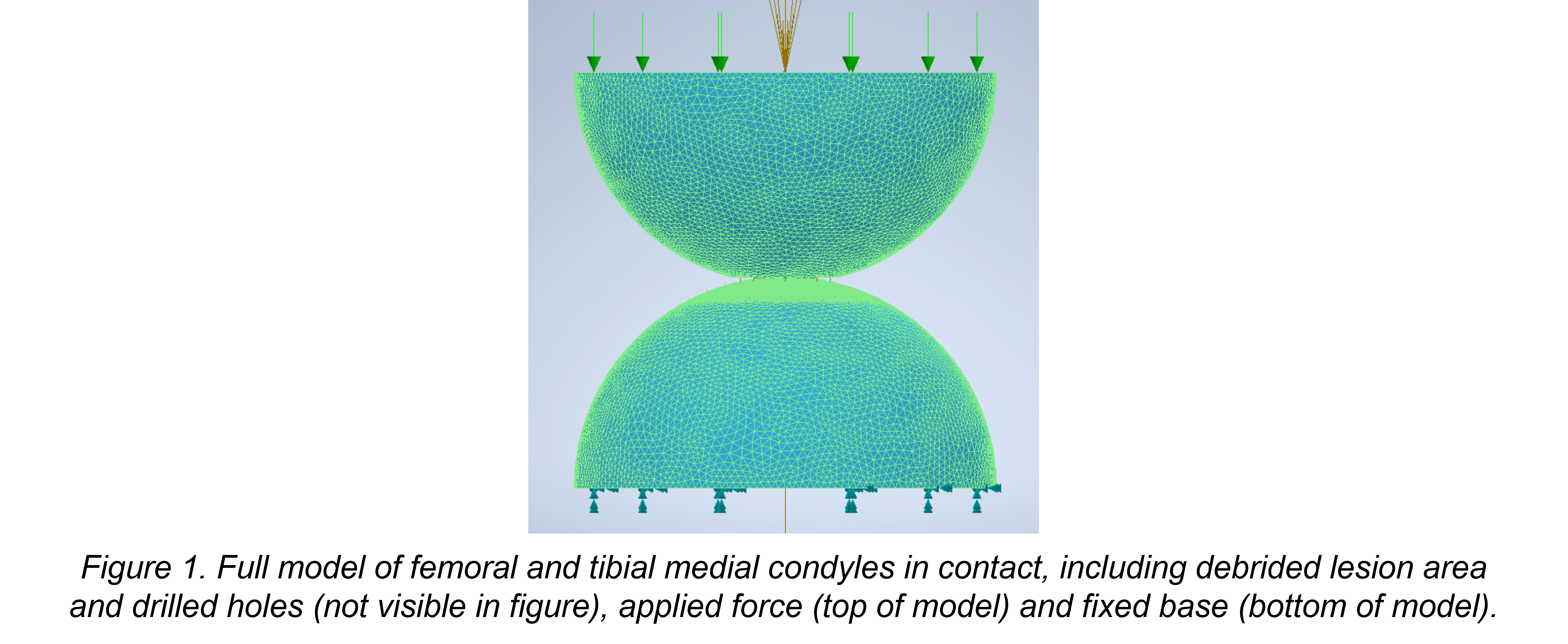
Computational experiments using finite element analysis were done using the values found from literature review to investigate the effects of hole coverage area and hole size on the stresses in a model of the femoral and tibial medial condyles in the knee (Figure 1). Holes of size 0.5mm, 1.0mm, 1.5mm and 2.0mm were simulated in separate models while keeping the coverage area at 5% to isolate the effect of hole size. Hole coverage area was then varied with values of 5%, 20% and 50% with 1.0mm holes to isolate effect of hole coverage area.
Results
It was found that the most consistent predictor for stresses in the condyles was the spacing between holes, as there is interdependence between hole coverage area, hole size, lesion area, hole spacing and hole number, with smaller spacings corresponding to higher stresses. The existing values for spacing in literature and in the clinic of 2-3mm is justified, as too small of spacing will result in higher peak stresses in the medial condyles. Thus, surgeons practising microdrilling or microfracture should stick to covering as much of the lesion with small holes of size 0.5mm or 1.0mm while keeping a spacing of 2-3mm to prevent breakage and high peak stresses as shown in the simulations.
The development of a microdrilling test rig was also continued with the implementation of a control system and user interface to an existing x-y table to be used with the microdrilling rig. This aims to improve ease of use, reliability, and precision. Test specimens are small, and thus the fine control of x and y displacement is crucial to perform more experiments to investigate drilling parameters and further optimise microdrilling.
Conclusions
Computational experiments that initially aimed to investigate hole arrangement by isolating hole coverage area and hole size with constant coverage area has shown that hole size, lesion size, coverage area, hole number, and spacing are interdependent, proving difficult to isolate any one parameter. However, it can be concluded that hole spacing plays an important role, and smaller spacing results in higher peak stresses. Further investigations should focus on isolating hole spacing, and further quantitatively optimising the arrangement of holes.
References
- KH. Pridie et al., “A method of resurfacing osteoarthritis knee joints,” Journal of Bone and Joint Surgery, 1959.
- EB. Hunziker et al., “An educational review of cartilage repair: precepts & practice – myths & misconceptions – progress & prospects,” Osteoarthritis and Cartilage, vol. 23, no. 3, pp. 334-350, 2015.
- L. Gao et al., “Subchondral drilling for articular cartilage repair: a systematic review of translational research,” Disease Models & Mechanisms, 2018.
* For more details please contact Dr Nan Li
| Project Type | Student Selected Component |
| Student | Shreya Singhal (School of Medicine) |
| Supervisor |
Dr James Ward (Engineering Department) |
| Advisor | Dr Sarah Greasley (Addenbrooke's Hospital) |
* For more details please contact Dr James Ward
| Project Type | Master of Engineering |
| Student | Stanislaw Tomaszczyk (Engineering Department) |
| Supervisor |
Prof Michael Sutcliffe (Engineering Department) |
Thesis Title
Mechanics of Human Bone - Lateral Hinge Fracture in High Tibial Osteotomy
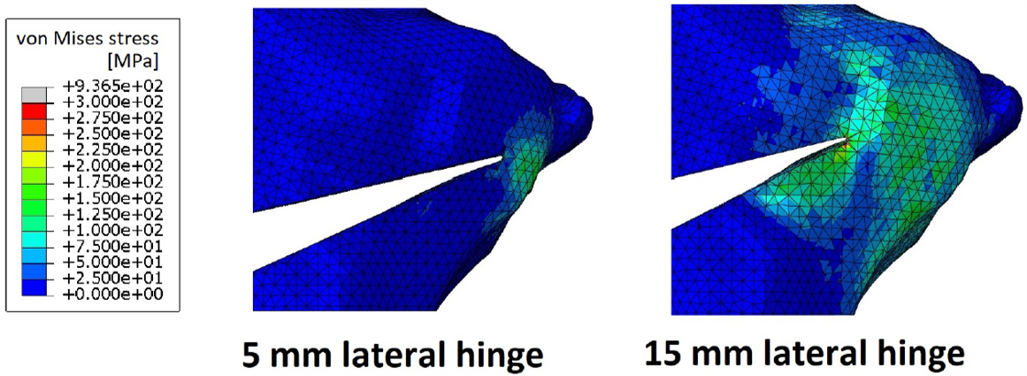
A comparison between the stress distributions for a 5 mm and a 15 mm lateral hinge at a 10 degree wedge opening
Abstract
Medial opening wedge high tibial osteotomy (MOWHTO) is a surgery aimed at correcting the varus deformity of the knee - one of the primary causes of knee osteoarthritis. It achieves this by adjusting the geometry of the proximal tibia. A cut is made through the bone using a surgical saw, it is then opened up by a desired angle and finally fixed in place using screws and a support plate. The surgery is known to be effective at reducing the loads acting on the medial compartment of the knee joint and therefore hindering the progression of osteoarthritis but it is also associated with multiple complications including lateral hinge fracture.
An investigation into the surgical guidelines for MOWHTO shows that the position of the apex of the cut is very poorly defined. It is clear that the position of the apex has a very big impact on the mechanics of the surgical process and therefore improving its positioning has a great potential for reducing the incidence of fracture. The aim of this project was therefore to investigate the impact changing the position of the apex has on the fracture risk using both experimentation and finite element modelling.
The main conclusion of this investigation was that, for a fixed applied displacement (i.e. fixed wedge opening), reducing the hinge width reduces the intra-operative fracture risk. However, robustness of the construct and faster healing process are factors that encourage wider hinges and they would have to be quantified in order to investigate the trade-off and find the optimum hinge width. Similarly, a risk analysis could be performed on the vertical position of the apex to understand the trade-off between the reducing the risk of the more dangerous type III fracture and reducing the overall fracture incidence. The observed correlation between the fracture nucleation
location and Takeuchi fracture type is a very useful result in that it allows to predict the failure mode without modelling the fracture event itself. Therefore, this study not only provides useful insight into the impact of the position of the osteotomy apex on the fracture risk in medial opening wedge high tibial osteotomy but also provides a strong basis for future work.
* Thesis Abstract and Contents (for more details please contact Prof Michael Sutcliffe)
| Project Type | Master of Science Thesis |
| Student | Tobia Nava (Master of Science in Engineering Design, KTH) |
| Supervisors |
Prof Michael Sutcliffe (Engineering Department, University of Cambridge) Prof Elena Farewik (Engineering Mechanics, KTH) |
| Advisor | Prof Andrew McCaskie (School of Clinical Medicine, University of Cambridge) |
Thesis Title
Surgical Microdrilling for Osteoarthritis Treatment - A Finite Element Study of Bone and Cartilage
Abstract
Surgical microdrilling is a potential treatment method for many patients suffering from osteoarthritis(OA). During microdrilling, the bone underneath the damaged cartilage is perforated to induce bleeding. The blood that enters the joint contains stem cells which help to restore the damaged cartilage. Microdrilling is known to affect the joint structure both biologically and mechanically, but knowledge regarding its structural effects is still limited. Based on literature review of previous research in microdrilling and adjacent areas, important influencing parameters were identified. Thereafter, a finite element study was performed simulating the bone and cartilage structures using a factorial design approach. Analysis of variance (ANOVA) was used to evaluate the results and to identify the relevance of the parameters: bone stiffness, bone thickness, cartilage thickness and microdrilling. The results revealed that the cartilage thickness is the most relevant factor regarding its influence on the mechanical properties of the joint, followed by bone thickness and microdrilling. Stresses in the bone can increase more than two times the value from the baseline. Based on the findings, the conclusion can be drawn that loss of cartilage has a significant influence on the contact mechanics of a joint. Furthermore, microdrilling affects the bone structure significantly, and alterations in bone thickness due to OA are not negligible. As a third element, an experimental test rig for studying microdrilling in mice was developed. The objectives of the development were to increase the user-friendliness and to enhance reproducibility. An actuated x-y table was designed including the corresponding control system using stepper motors and a Raspberry Pi.
* Thesis Abstract and Contents (for more details please contact Prof Michael Sutcliffe)